Tuesday, October 9, 2012

Unknowns Project

This is still under construction!

One of our goals for this semester was to isolate and identify 3 unknown fungi (unknown to us, anyway). There is a list of common fungi that we cannot turn in, but we can collect from pretty much anywhere for this project. I work with endophytic entomopathogenic fungi in cotton targeting thrips and spider mites, and so I was curious if these arthropods harbor any fungi in heathy conditions that I should know about when doing work like this. I was also curious if they share similar fungi as the food they feed on. In this case, my colonies feed on excised bean leaves, so I did a comparison of endophytes in freshly cut bean leaves and leaves that had been fed upon for one week. The specific groups that I plated out to look for fungi were:

  1. Surface sterilized adult spider mite females
  2. Non surface sterilized adult spider mite females
  3. Surface sterilized adult female thrips
  4. Non surface sterilized adult female thrips
  5. Surface sterilized immature thrips
  6. Non surface sterilized immature thrips
  7. Freshly cut surface sterilized bean leaves
  8. One week old surface sterilized bean leaves
Adult female Western Flower Thrips.

Two-spotted spider mite adult male. 

To surface sterilize the spider mites and thrips I placed 10 individuals in 70% ethanol for 1 minute, removed them and placed them into 2% bleach for 1 minute, and then removed them into a sterile water wash. The sterilized individuals were then placed evenly throughout a water agar plate. Each group was plated separately, i.e. thrips were not plated with spider mites.

Surface sterilization of plant material followed the same order and dilution, however, leaves remained in ethanol for 2 minutes and in bleach for 3 minutes. Leaves were placed directly on a water agar plate, and with a sterile scalpel, cut into sections to expose more internal surface area to the agar.

The non sterilized mites and thrips were simply pulled from the colony with a sterile tool and placed on a clean agar plate. All of this work was done under a sterile laminar flow hood on September 27, 2012. Once plated, the petri dishes were sealed with parafilm and placed at room temperature near a window to simulate a natural photoperiod.

The laminar flow hood where I did my work, along with surface sterilization materials, a thrips colony and a fresh bean plant.  
Adult female thrips in a beaker containing 70% ethanol. 
A series of beakers for surface sterilizations. From back to front: 70% ethanol, 2% bleach, sterile water.

Adult female thrips across a water agar plate.


A "condo" containing a thrips colony that has been feeding for one week.
Previously fed upon leaves undergoing their surface sterilization.


Adult female spider mites on their water agar plate. I promise they are there, you just have to look closely.

Fresh bean leaves on water agar that were cut to expose internal leaf tissue to the agar.

After six days, the only plates that had growth were the non sterilized adult female mites, the non sterilized female thrips, and the one-week-old bean leaves. 

From the plates that showed growth, agar plugs were removed from areas with clean boundaries that did not visibly have contamination. They were transfered under the flame of a bunsen burner to new, clean 1/2 PDA plates.

A spider mite showing fungal growth.

What appears to be a thrips egg showing fungal growth.

At one week on the 1/2 PDA, there appears to be three distinct fungi growing in my plates. The used leaf and spider mite palates each have a unique fungus, but the thrips plate appears to have a fungus that has also shown up on the spider mite plate. When I get time, I will put these on slides under the microscope to identify what fungi I might have. I have a feeling that they may be on the common list, so I better get to finding new sources of fungus!

Fungi growing from non sterilized thrips.

Two types of fungi from non sterilized spider mites.

Previously fed upon bean leaf on 1/2 PDA growing an unknown fungus.


October 3 Lab

The goals for this lab were to:
  • Familiarize ourselves with some of the Hyphomycetes
  • Check in on our Neurospora crosses from last week
  • Check in on our Ustilago infected corn from last week and stain for the fungus

The Hyphomycetes
The hyphomycetes are a broad term for conidiating fungi. These are very common and it would be a good idea to become familiar with the group in general. We looked at 12 different species throughout the lab to get a better idea of their spore shapes, hyphal forms, conidiophores and other various structures. I drew pictures of each of these fungi, and took a few photographs of some. The following images were observed from either a tape or squash mount slide. 

Trichoderma viride showing a conidiophore on the right, and after looking at the Illustrated Genera of Imperfect Fungi (IGIF, from here on out), what I can only assume is hyphae layered with conidia on the left and bottom. 

The coolest conidia we looked at today. Pestalotia conidia that bear appendages on both sides.  Hyphae threaded together on the right.

Fusarium graminearum conidia. They remind me of compartmentalized bananas. 

Monilinia fructicola. This looks like a section of the branched conidiophore on top, loose conidia in the center and an incompletely drawn hypha on bottom. IGIF shows a much better image.

Rhizoctonia solani does not have conidia or asesexual fruit bodies. My sample appears to be a bit dried out, but similar in appearance to IGIF. 

Thielaviopsis brasicola showing its aleuriospores. This also produces phialospores, but they are not shown here.
Aleuriospores of Thielaviopsis brasicola. 

Epicoccum spp. The conidia did not remain with the conidiophore in my images.
Microscopic image of Epicoccum conidia.


This Curvularia spp. reminds me of alternaria, except these are a more grey conidia. The conidia can be straight or bent under the scope.

Not the best Aspergillus niger mount that I have done. You can see conidia in chains or part of the conidiophore here. 

Botrytis cinera. The only useful part of this image is my attempt to demonstrate the slender nature of this fungus. A better image can be seen in IGIF.

Alternaria brassicola. We have seen this before, but here again is a simple drawing. The conidia are brown.

We were also going to look at Colletotrichum coccodes and Nigrospora, but they were contaminated with an unknown fungus. 

Neurospora crosses from last week

We each checked in on our Neurospora crosses from last week to see if any perithecia containing ascospores had formed. You can see growth on my plate, and you can see were the two fungi have met in the center of the plate. Interestingly enough, my plate did not bear perithecia in the center of the plate, but instead on the far ends of the plate. They were not completely developed this week, but I will try to get a better image next week.
My Neurospora cross one week after plating. 
Ustilago infected corn 

Last week we infected one-week-old corn plants with Ustilago maydis. This fungus causes corn smut and leads to large, bulbous growth on the plant after affecting plant cell growth and division. After just one week there were already visible signs of the disease.  


Damage present on the corn plant after just one week.
A better view of a small gall forming on one of my infected plants from last week. 
To confirm that a fungus was causing these galls, I removed a portion of the plant and began a staining protocol that binds a blue dye to the chitin in the fungal cell wall. To begin, I placed the excised leaf material in a plastic petri dish and submerged this leaf in a 2:1 Acetic acid: ethanol solution to clear the leaf. The leaf remained in this solution for 24 hours, was then removed from the solution, rinsed with ethanol, and placed in a microcentrifuge tube with lactophenol blue. This solution has three purposes. It kills any living specimens, preserves fungal components, and stains chitin blue. I kept the leaf in this solution for 4 hours, and then removed it into a jar of ethanol to remove any excess blue dye. I kept the leaf in this solution for several hours, then placed the leaf onto a slide, covered with a long cover slip, and then observed the locations of fungus in the plant.

The excised leaf material freshly placed in acetic acid/ethanol mix.
My stained corn leaf showing pockets of blue where the fungus is present.

The color is a bit off on my camera, but you can see blue color has remained in the veins of the plant. 

The location of a gall. You can see a congregation of blue mycelia.

A small blue lesion, likely the start of a gall.

Closed stomata on the underside of the leaf. You can see the cells dotted with blue spots,  spores perhaps?


September 26 Lab

The goals for this lab were to:

  • Observe ascospores, microconidia, and macroconidia on Neurospora
  • Cross Neurospora strains
  • Work with the Basidiomycete Ustilago maydis to infect health corn plants, learn the life cycle, and begin to observe overtime the symptoms that appear on infected corn plants
  • Look at dimorphism in the Zygomycete, Mucor rouxii
Ascospores, microconidia and macroconidia of Neurospora

Three types of media were used to produce each of the spore types. A nutrient-poor medium in a petri dish produced microconidia, nutrient-rich medium in a petri dish produced macroconidia, and media contained in a microcentrifuge tube produced the ascospores. To collect macroconidia, I gently scraped a small portion of the growing fungus from the agar and placed this on a slide with a water drop. A cover slip was placed on top of this. You can see the macroconidia in the following picture.

Neurospora macroconidia
To collect the ascospores, I used a sterile loop that had been dipped in sterile water. I then took this look and gently collected material that I thought would have spores. This was placed on a slide with a drop of water in the center, and then a coverslip was placed on top of this. 

Neurospora ascospores.
I was not able to get a good picture of the Neurospora microconidia, and my drawing does not provide a lot of detail. Hopefully someone else in the lab will have a picture on their blog.
Crossing of Neurospora strains

There were several strains of Neurospora available in the lab to cross. I chose SMRP11 and SMRP10 which are two opposing mating types. To do this cross, I simply took (under sterile conditions) an agar plug from each of the stock plates and then placed them on opposite ends of a clean petri dish with SC solid media. I sealed the dish in parafilm and then place this in a clear plastic box in the back of the lab. I will look at this next week to see if I have had a successful cross. 
What my Neurospora cross looks like immediately after the agar plugs were placed in the plate.
 Dimorphism in the Zygomycete, Mucor rouxii

In this lab we attempted to observe dimorphism of Mucor rouxii, which under aerobic conditions takes on a hyphal form and when under anaerobic conditions takes on a yeast form. Small cups with media had been previously inoculated with this fungus for our use. A small sliver of agar was cut from the center of the cup, making sure to include agar that both had access to oxygen on the surface, but that also lacked oxygen from the bottom of the cup. Ideally you should be able to see a gradient from top to bottom of the two forms, however I found that I needed to pick and choose through the top to get hyphal images, as well as from the bottom for yeast forms. Eventually I was able to successfully get a few images to demonstrate this dimorphism.


Mucor rouxii hyphal form taken from near the surface of the agar.

If you look closely near the cut-off end of this image you can see the formation of the yeast type cells of Mucor rouxii.
Also in the Mucor cups I was able to see many of these round, brown sporangiophores. 

Infection of corn plants with Ustilago maydis

To learn more about the infection process and life cycle of a Basidiomycete, we began today the infection process of corn by Ustilago maydis. This fungus causes corn smut, which will create galls on the leaves and reduce yields. To infect our clean, healthy plants that were one week old when provided to us, we simply injected Ustilago maydis into the stem of the plant until the solution began to flow from the whorl of the plant. I infected two plants, and I was able to inject about .5 mL spore solution per plant.

Here is an example of an injection of Ustilago maydis into a one-week-old corn plant stem. 
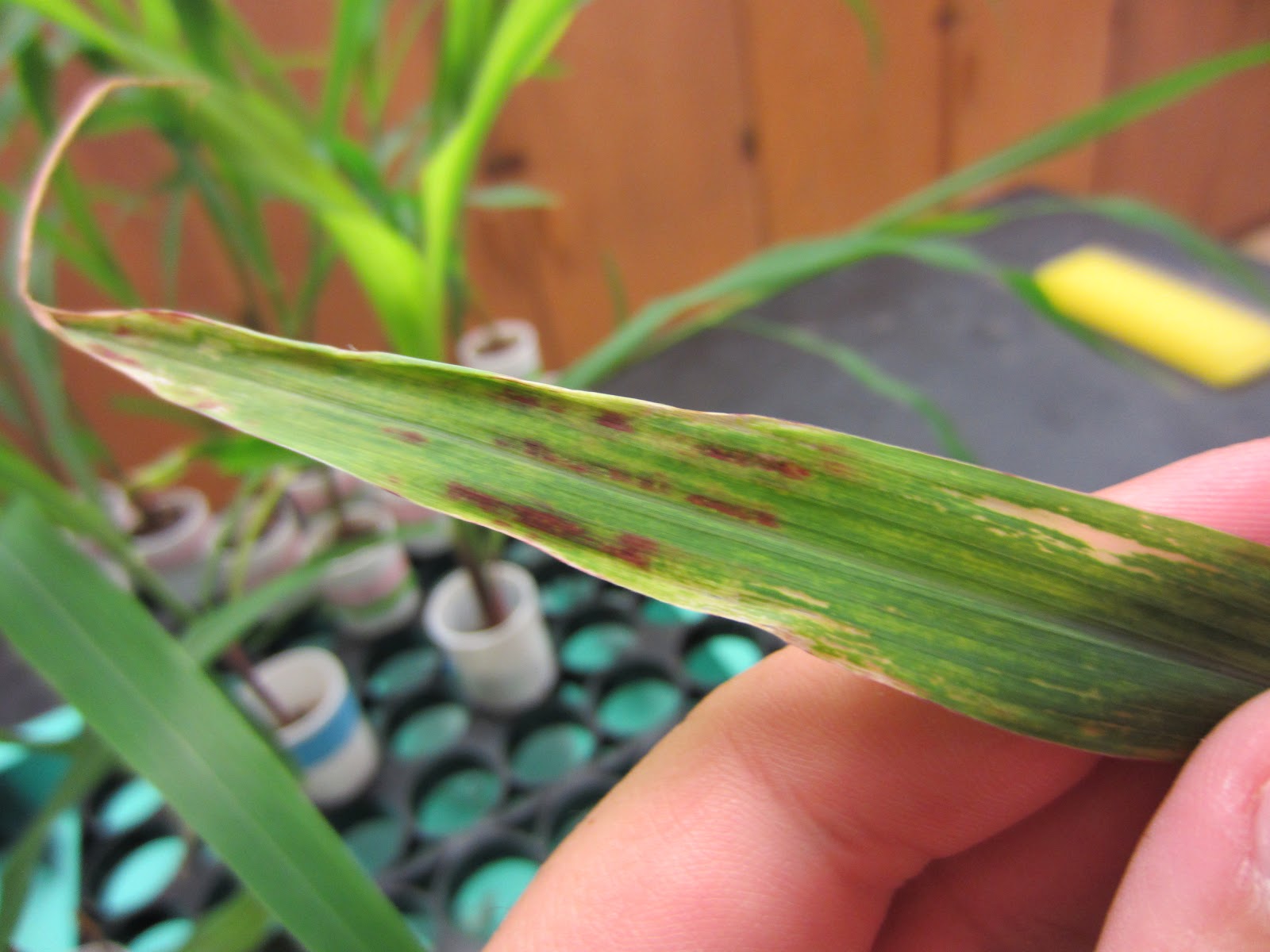
There were also two-week-old plants that had been infected one week prior for our viewing pleasure. You can already see galls forming on several of the leaves.

From a distance, this plant appears to be a healthy, two-week-old corn plant. 

Upon closer examination, reddening and damage are more obvious in this plant.

Here you can clearly see small galls forming from and infection of Ustilago maydis.


Monday, October 8, 2012

September 19th Lab

The goals for this lab were to:
  • Look at various fungi that make zoospores
  • Observe Saprolegnia cultures that were plated over time
  • Culture Allomyces from previously baited seeds
  • Observe various life cycle videos
We were also going to cross Neurospora, but due to time constraints this was left to do the following week.

Fungi that make zoospores:

Initially we were supposed to look at Allomyces, but the culture did not grow so instead we looked at the Oomycte, Phytophthora. Phytophthora is not considered a fungus, but it is similar to Allomyces in that it produces zoospores. They are considered protists, and although they can cause disease, look and smell like fungi, they differ from fungi in their cell wall structure as well as having differing sequence data. Today we specifically looked at Phytophthora infestans the causal agent of Potato Blight. Unfortunately photos do not do a very good job of showing the motile zoospore, but I was able to capture some rough footage of a zoospore in action.

If you look closely in this video you can see the flagellum on this zoospore. 

Saprolegnia transfers from September 9, 12, and 18

The next thing we looked at were a series of Saprolegnia transfers that were plated on September 9, 12, and 18. It should be noted that Saprolegnia is also not a true fungus, it is instead another Oomycete. To better view the specimen I used a method Dr. Shaw had told us about in a previous lab . The goal of this method is to elevate the cover slip from the specimen so that it does not crush the sample or reduce any movement. To do this, I broke an additional cover slip and placed some fragments on a microscope slide, placed my sample in between the fragments, and then placed the whole cover slip over the entire mount. Here is what it looks like:
A simple way to elevate your cover slip is to place fragments in between the slide and the cover slip. 


Here is my drawing from the oldest (10 days old) Saprolegnia transfer. You can see sporangia and some free spores, as well as rhizoid like growth. 
10 day old Saprolegnia sample

The seven day old Saprolegnia was much better for me to see the small, motile zoospores. In this video, you can occasionally see the zoospores moving in tight circles or back and forth. 


I could not see anything from the one day old transfer. This is apparently not enough time, or I did not have enough skill, to properly see this sample. 

Culture Allomyces from baited seeds

In the lab there were seeds available that had been previously baited with Allomyces. The goal was for us to be able to somehow obtain some of the Allomyces from the seed, transfer it to an agar plate, and attempt to isolate and grow Allomyces for ourselves. I looked at my seed under the dissecting microscope and was able to see movement in the water, but no obvious zoospores. I pipetted 1 mL solution from around the seed and then transfered this to a clean PDA plate. I then sealed this plate with parafilm, took it back to my lab, and set it out to see if I could get any growth. Unfortunately, all that I was able to culture on my plate was bacteria. I will have to find a better method for transferring the fungus in question next time I try this.


Monday, September 24, 2012

September 12 Lab

The goals for this lab were to cover:
  • Subculturing 
  • Aspergillus cultures
  • Riddell mounts
  • Hyphal growth
  • Media Variants
For subculturing, you want to select the appropriate media (more on this later) and under sterile conditions, transfer a small section of previously plated fungus on to the new media. This is a pretty simple procedure, and this will be necessary when isolating fungi for our unknowns.

We also looked at a number of Aspergillus species to see if we could tell the difference in fungi of similar structure. This group has simple, round spores that make identification difficult unless the intact conidiophore is viewed. This turned out to be fairly difficult for me to tell the difference, but I think as I get better at preparing slides to look at my specimens, this will become easier. Here are some drawings of the various samples we looked at.
Aspergillus sojae, squash mount.

Aspergillus flavus (atoxigenic strain), squash mount

Aspergillus oryzae, agar block.

Aspergillus paraciticus, squash mount.

Aspergillus nidulans, squash mount

Aspergillus niger, squash mount

Aspergillus tamari,  squash mount


     The Riddell mount is another technique we learned in the quest to better view conidiophores. The classical version of this technique is to place a cut piece of agar on to a microscope slide that will be placed in a clean petri dish on top of a bent glass rod. The glass rod elevates the slide so that water can be added to the petri dish to avoid letting the agar dry out. From here, the fungus in question will be transfered to the sides of the cut agar block, and then a cover slip will be placed on top of this agar block. After a few days, as the fungus begins to grow, some growth should come in contact with the cover slip. This cover slip can then be transfered to a new, clean slide with a drop of water and be viewed under the scope. It is ideal to make several of these mounts that can be viewed over the course of a few days to catch the optimal stage in growth for viewing. There is a modified version to these mounts that requires less supplies and time. Simply take a clean agar plate, cut a few blocks from the edge, and then place these blocks on the side of the plate that has uncut agar. This method will help to  keep moisture in the agar block.

     Finally, we also learned about various types of media that we can use to grow our various fungi with. The best media may be different for each fungus and the use of several types may be necessary to find the right media to properly grow a particular fungus. More on the various types of media can be found here:

http://classes.midlandstech.com/carterp/Courses/bio225/chap12/fungalculturemedia.htm


Thursday, September 13, 2012

Lab 1...A bit late

     I didn't get a chance to post about last week's lab (September 5) but I will go ahead and talk about it now. After going through everyone's blogs, we covered setting the proper Kohler illumination on our compound microscopes to better view specimens. Proper set-up will help to achieve clear, contrasting images that are evenly lit. 

    We also got a chance to practice making squash mounts and tape mounts of Thielaviopsis brassicola, Cladosporium, Aspergillus niger, Alternaria brassicicola, and Pythium ultimum

    Before reviewing these two mount techniques, let's first cover how we are maintaining a sterile field. Let's also assume for any subsequent work, unless stated otherwise, working in a sterile field will refer to this technique. Simply begin by setting up and lighting a Bunsen burner on a level surface. Convection produced from the flame will keep any petri dishes in close proximity to the base of the burner in a clean field. When opening a petri dish, be sure to open towards the flame, to avoid any possible contamination from breathing on the plate. Also, be sure to pass any tools that will come in contact with the agar, petri dish, or fungus, through the flame for a few seconds to sterilize them before use.   
Convection created by the flame will keep your petri dish in a clean environment. Photo taken from http://www.stbedes.ngfl.ac.uk/curriculum/science/microbi.htm
Squash mount: In a sterile field, place a drop of sterile water on a glass slide. Using a scalpel, probe or loop, retrieve a small mass of fungus from a previously inoculated plate. If agar is collected with this sample, try to keep it to a minimum to make it easier to view the finished mount. The sample should be placed in the water droplet on the slide, and a clean cover slip should be placed over the center of the sample. Using the flat end of your probe, gently press down on the cover slip to flatten and even the sample to better view under the scope. This technique takes some practice and is greatly dependent on the particular fungus. Thankfully I will get lots of practice with this method this semester and should get better with time.


     Tape mount: This method (if you get good enough at it) can be used to see conidiophores that may be damaged or otherwise not visible in a squash mount. Basically you take a small piece of tape and very gently touch the tape to the plated fungus. This tape can then be placed on to a drop of water on a clean slide. No cover slip is necessary for viewing under the microscope. While I was able to see different structures with this method, I still prefer the squash mount. 
An example of a squash mount. Clearly I still need practice with this method. 

   Use of the Hemocytometer for counting cells was taught during this lab as well. I have used this technique before when making spore solutions to inoculate cotton seeds with fungal endophytes. The basic gist is to take an aliquot of spore solution and place them on to a specialized gridded slide that aides in counting the number of cells per square on the grid. You can then do the math to figure out how many cells you have per volume in your solution. Using a particular dye you can also determine which cells are alive and which are dead.
This is an example of what the entire grid on the hemocytometer  looks like. The  circled squares are where my lab tend to take counts from.
    I didn't take any pictures from the microscope of the fungi we looked at, but I was able to draw some representative pictures...

Thielaviopsis brassicola squash and tape mounts. Both techniques let me see similar structures.  
Cladosporium sp. squash mount and tape mount drawings. I was able to see more structures with the tape mount that I could not capture with my squash mount.
Aspergillus niger squash and tape mounts. I was only able to see spore chains in my tape mount.
Alternaria brassicola squash mount only. The conidia were easily seen with this method.
Pythium ultimum squash mount. There were a lot of unknown structures, but we believe that the thin-walled cells are young oogonium and the thick-walled are developing cells after fertilization.